Fluorescent proteins (FPs) have revolutionized the field of cellular and molecular biology, enabling advanced imaging techniques that were once unimaginable. These proteins have become invaluable tools for studying cellular structures, dynamics, and functions in real time, especially through live-cell imaging. They are integral to fields such as cell biology, developmental biology, and cancer research.
What Are Fluorescent Proteins?
Fluorescent proteins are proteins that absorb light at one wavelength and emit it at another, producing bright, colored light. These proteins come in a variety of colors, including green, red, blue, and cyan, making them highly versatile for studying various aspects of cellular activity simultaneously. The discovery of the green fluorescent protein (GFP) from Aequorea victoria was a breakthrough, and since then, many variants have been developed with different excitation and emission spectra.
How Fluorescent Proteins Are Used in Advanced Imaging Techniques
Fluorescent proteins are key players in modern imaging techniques such as confocal microscopy, live-cell imaging, and super-resolution microscopy. They allow researchers to visualize specific proteins, cellular structures, or even entire cells within living organisms, without the need for invasive techniques. Here’s how they are used in different imaging techniques:
- Live-cell imaging: Fluorescent proteins enable scientists to track dynamic biological processes in live cells, such as protein localization, cell division, and intracellular trafficking. These proteins can be expressed in specific cellular compartments, allowing for real-time monitoring of cellular processes.
- Microscopy: Advanced microscopy techniques, including confocal and fluorescence microscopy, rely on fluorescent proteins to provide high-resolution images of cellular structures. Fluorescent proteins provide an unmatched combination of spatial and temporal resolution, enabling detailed visualization of the fine details within cells.
- Super-resolution microscopy: Techniques like STORM (Stochastic Optical Reconstruction Microscopy) and PALM (Photoactivated Localization Microscopy) use fluorescent proteins to break the diffraction limit of light and achieve nanoscale resolution. These techniques are widely used to study complex protein-protein interactions and cellular structures at unprecedented resolution.
Key Applications in Research
Fluorescent proteins have broad applications in both basic and applied research:
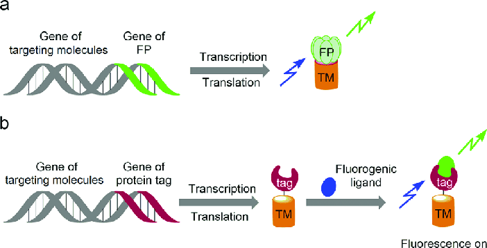
- Protein tracking: Fluorescent proteins are commonly fused to other proteins to study their localization and dynamics. This is particularly useful in studying protein-protein interactions, signaling pathways, and cellular responses to stimuli.
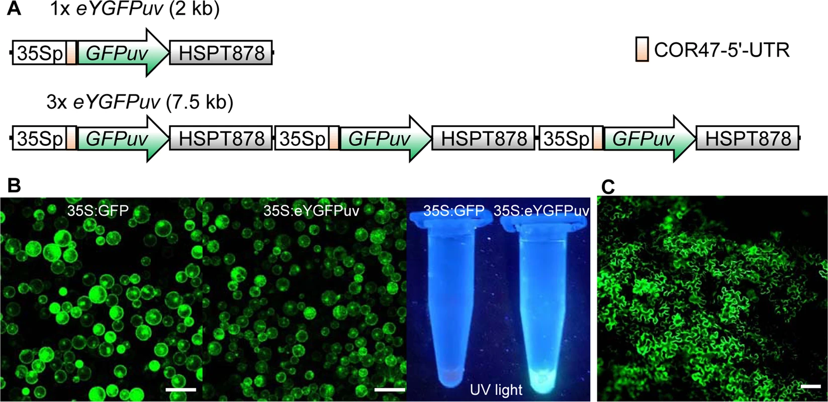
- Gene expression analysis: Researchers use fluorescent proteins to study gene expression by linking them to promoters of interest. By measuring the fluorescence intensity, scientists can monitor the activity of specific genes in real time.
- Cancer research: In cancer biology, fluorescent proteins have been used to track tumor progression, metastasis, and drug responses. By labeling tumor cells with different fluorescent proteins, researchers can distinguish between tumor and normal cells in live animals, providing insights into tumor behavior and response to treatment.
Popular Fluorescent Proteins and Reagents
Various types of fluorescent proteins are available for different applications. Some of the most popular include:
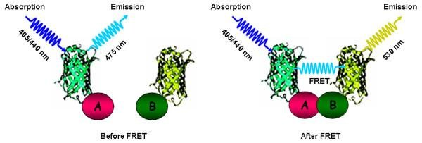
- Cyan Fluorescent Protein (CFP): Often used in dual-color imaging and FRET (Förster Resonance Energy Transfer) studies. The Cyan Fluorescent Protein is available at Gentaur, providing high brightness and excellent photostability for a range of imaging applications.
- Green Fluorescent Protein (GFP): The most widely used fluorescent protein, GFP is ideal for live-cell imaging and gene expression studies.
- Yellow and Red Fluorescent Proteins: These proteins are used for multicolor imaging, enabling simultaneous tracking of multiple cellular components in different colors.
Fluorescent Protein Reagents
To enhance the utility of fluorescent proteins, various reagents are available that can optimize imaging, improve signal-to-noise ratios, and facilitate better protein expression. These reagents include:
- Fluorescent Protein Tagging Kits: These kits provide everything needed to tag proteins with fluorescent markers, allowing for easy integration into live-cell studies.
- Fluorescent Dyes: Used in conjunction with fluorescent proteins, these dyes enhance the visibility and contrast of labeled cellular components.
For example, Cyan Fluorescent Protein reagents from Gentaur can be incorporated into various imaging workflows to improve cellular fluorescence.
Conclusion
Fluorescent proteins have undeniably transformed the field of cellular imaging and research, enabling scientists to explore the inner workings of cells in live organisms. With the continuous advancement of fluorescent protein technology, the possibilities for discovery and innovation in biology are virtually limitless. Whether you are conducting protein localization studies or investigating disease mechanisms, fluorescent proteins are an essential tool in every researcher’s toolkit.
![Examples of alpha herpesvirus research using fluorescent proteins and biosensors. (A) Three pseudorabies virus (PRV) strains expressing mCherry (red), enhanced yellow fluorescent protein (EYFP; depicted in green), or mCerulean (cyan) segregate to produce single-color plaques after spread from neurons [35]; (B) PRV expressing pHluorin, a pH sensitive FP biosensor (green), and mRFP1 fused to viral capsid protein VP26 (red), reveals the exocytosis of single virus particles [36]; (C) PRV expressing mRFP1 fused to viral capsid protein VP26 (red) co-transport with mCitrine-tagged kinesin-3 microtubule motors (green) in neuronal axons. Kymograph reveals movement of individual virus particles over time [37]; (D) PRV expressing GCaMP3, a Ca2+ biosensor, reveals synchronized firing of infected neurons in vivo [31]; (E) An explanted peripheral nervous system ganglion infected with a mixture of three PRV strains expressing mCherry (red), EYFP (green), or mCerulean (cyan) [38]. All images are reproduced with permission of original authors. Examples of alpha herpesvirus research using fluorescent proteins and biosensors. (A) Three pseudorabies virus (PRV) strains expressing mCherry (red), enhanced yellow fluorescent protein (EYFP; depicted in green), or mCerulean (cyan) segregate to produce single-color plaques after spread from neurons [35]; (B) PRV expressing pHluorin, a pH sensitive FP biosensor (green), and mRFP1 fused to viral capsid protein VP26 (red), reveals the exocytosis of single virus particles [36]; (C) PRV expressing mRFP1 fused to viral capsid protein VP26 (red) co-transport with mCitrine-tagged kinesin-3 microtubule motors (green) in neuronal axons. Kymograph reveals movement of individual virus particles over time [37]; (D) PRV expressing GCaMP3, a Ca2+ biosensor, reveals synchronized firing of infected neurons in vivo [31]; (E) An explanted peripheral nervous system ganglion infected with a mixture of three PRV strains expressing mCherry (red), EYFP (green), or mCerulean (cyan) [38]. All images are reproduced with permission of original authors.](/web/image/179427-e8df07c7/image.png?access_token=6a3dfa68-1a5f-43b3-9a74-db9c765e40fb)


